POCUS-Guided JVP Assessment Study Protocol Training

Point-of-Care Ultrasound for Heart Failure Management
Anatomical Reference
00:00

Forms

✓ Checkout Form
✓ Demographics
✓ Data Collection
Butterfly iQ step screenshot

Study Protocol Steps

Score: 0/12

💡 Protocol Tip

Follow the numbered sequence carefully. Each step is critical for study compliance.

Protocol Feedback

Completed Steps

    No steps completed yet.

    ×

    POCUS Protocol Test Complete!

    Your test results have been encoded into a verification code. Please send this code to the study administrator.

    📧 Email Results to Administrator

    You can also copy the code above and paste it into an email manually.

    ×

    POCUS JVP Study - Terms & Procedures

    POCUS-GUIDE-HF: Study Overview

    Title: POCUS-GUIDE-HF: Point-of-Care Ultrasound-Guided Diuresis in Acute Decompensated Heart Failure to Reduce 30-Day Readmissions and Prevent Acute Kidney Injury

    Background: Heart failure (HF) hospitalizations are a major contributor to healthcare burden, with high rates of 30-day readmissions and associated acute kidney injury (AKI). Accurate assessment of volume status is critical in optimizing diuretic therapy, yet traditional clinical evaluation is often unreliable.

    Objective: This study aims to evaluate whether point-of-care ultrasound (POCUS)-guided diuretic management can reduce 30-day hospital readmissions and prevent AKI in patients hospitalized with acute decompensated heart failure (ADHF).

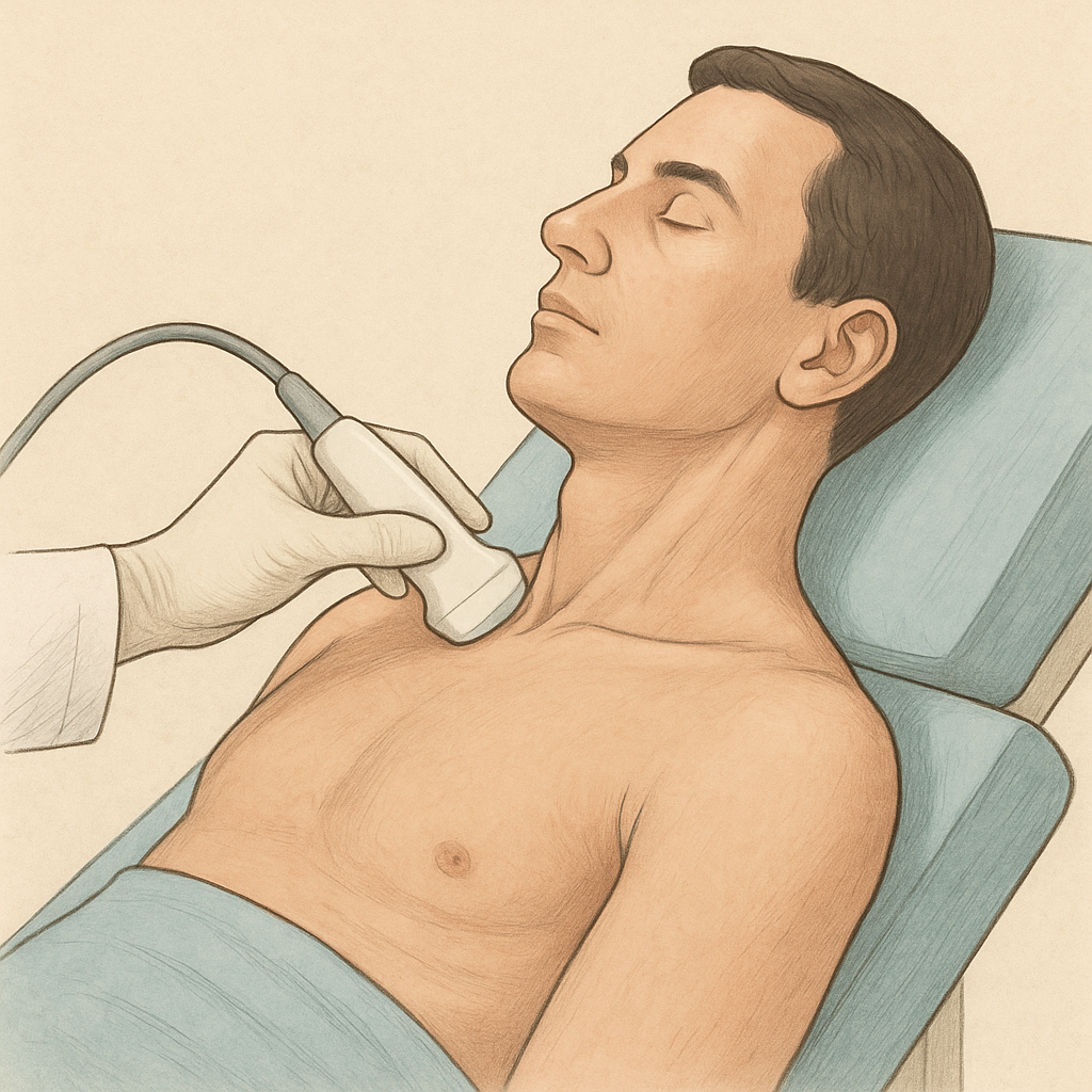
    Study Design: A prospective, randomized, controlled trial comparing POCUS-guided diuretic management to standard care in patients hospitalized for ADHF. POCUS will be used to assess right internal jugular vein (RIJV) collapsibility during the Valsalva maneuver, guiding diuretic adjustments. A distensibility index > 66% generally signals euvolemic - higher percent lower volume. Under 66% signals volume up.

    In the POCUS group, daily ultrasound findings will be communicated to the clinical team with specific recommendations for diuretic adjustments. We will also evaluate adherence to these recommendations. In the control group, POCUS assessments will be performed daily for research purposes, but findings will not be shared with the clinical team.

    Inclusion Criteria:
    • Age ≥18 years
    • Hospitalized with a primary diagnosis of ADHF
    • Planned treatment with IV loop diuretics
    • Ability to perform and tolerate the Valsalva maneuver
    Exclusion Criteria:
    • Presence of a left ventricular assist device
    • Significant anatomical limitations precluding adequate IJV assessment (e.g., neck deformities, tracheostomy)
    • Known metastatic cancer or limited life expectancy <6 months
    • Severe tricuspid regurgitation or congenital heart disease affecting venous return
    • Hemodynamic instability requiring vasopressors or mechanical circulatory support
    Primary Outcomes:
    • 30-day all-cause readmissions
    • Incidence of AKI during hospitalization
    Secondary Outcomes:
    • Changes in quality of life (KCCQ scores)
    • Length of hospital stay
    • Time to transition to oral diuretics
    • Adherence to POCUS-based recommendations in the intervention arm

    Significance: By incorporating POCUS into clinical decision-making, this study seeks to improve patient outcomes, reduce healthcare costs, and provide a scalable model for HF management across diverse healthcare settings.

    Funding: This project is supported by a Postdoctoral Fellowship Award from the American Heart Association.

    Possible patient-friendly explanation: “We’ll use a quick bedside ultrasound of your neck veins each day to see how much excess fluid you have (in addition to your weight, exam and labs), aiming to keep fluid off your lungs without taking too much fluid."

    Video Overview: 60-Second Scan Protocol

    This video demonstrates the complete 60-second scanning procedure, including patient positioning, probe placement, and the timing of the three Valsalva maneuvers.

    Key Timestamps:
    - 0-15s: Baseline IJV capture.
    - 15s, 30s, 45s: Patient performs Valsalva maneuver.
    - 60s: Scan concludes.

    Image Sending Protocol (Text Guide)

    Follow these steps within the Butterfly iQ+ App to finalize and share your scan:

    1. Select Vascular Access preset.
    2. Perform a one-minute capture.
    3. Tap Saved to review the clip.
    4. Open Patient Details.
    5. Enter Hospital and Room #.
    6. Save as Draft.
    7. Go to Studies and open the most recent draft.
    8. Email co-investigator.
    POCUS (Point-of-Care Ultrasound)

    Bedside ultrasound performed by clinicians at the patient's location for immediate diagnostic information. In this study:

    • Uses high-frequency linear probe (5-10 MHz)
    • Provides real-time vessel visualization
    • Non-invasive alternative to invasive monitoring
    • Allows dynamic assessment during maneuvers
    Internal Jugular Vein (IJV)

    Major venous structure in the neck used for JVP assessment:

    • Located lateral to carotid artery
    • Directly reflects right atrial pressure
    • Easily compressible (unlike artery)
    • Shows respiratory variation
    • Diameter changes with fluid status

    Study measures IJV diameter at multiple time points to track fluid removal effectiveness.

    See also: Hypervolemic IJV appearance | Euvolemic IJV appearance

    Valsalva Maneuver - Standardized Protocol

    Forced expiration against closed airway to increase intrathoracic pressure:

    • Exact patient instruction: "Take a deep breath, then hold it and bear down like you're having a bowel movement"
    • Timing in study: Performed at 15, 30, and 45 seconds during 60-second scan
    • Expected response: IJV distension during strain phase (may increase 2-3x baseline)
    • Safety: Monitor for dizziness, lightheadedness, or discomfort

    Use exact wording for consistency across all study participants!

    Jugular Venous Pressure (JVP)

    Indirect measurement of right atrial pressure:

    • Traditional method: Visual inspection (often inaccurate)
    • POCUS method: Precise IJV diameter measurement
    • Normal: 6-8 cm H₂O
    • Elevated in heart failure due to fluid overload
    • Decreases with effective diuresis
    Study Randomization

    Participants randomly assigned to one of two groups:

    • POCUS Group: Ultrasound findings shared with clinical team
    • Control Group: Ultrasound performed but findings NOT shared
    • Stratified by age, gender, and medical history
    • Ensures comparable groups for valid results

    Study team does NOT make treatment decisions - only provides data to clinical team.

    Demographics for Stratification

    Key data collected for randomization using the Demographics Form:

    • Age and gender
    • Ejection fraction (if known)
    • Baseline creatinine
    • Diuretic dose
    • Length of stay
    • Comorbidities (diabetes, hypertension, CKD)
    Omoclavicular Triangle & Probe Positioning
  1. Select on Butterfly App Presets: Carotid
  2. Set head of bed elevation to 45 degrees
  3. Anatomical landmark for IJV visualization:

    • Bounded by sternal and clavicular heads of SCM muscle and clavicle
    • IJV runs lateral to carotid artery in this space
    • Probe orientation: Point blue dot toward patient's posterior neck
    • CRITICAL: Use LIGHT TOUCH - vessel easily compressed
    • IJV appears larger, more lateral, and compressible vs carotid

    Remember: Dot to posterior neck, light touch technique!

    Patient positioned at 45 degrees with POCUS probe applied to the omoclavicular triangle.
    Daily Assessment Protocol

    Study requires daily ultrasound until discharge:

    • Same time each day when possible
    • Document any changes in clinical status in your progress note
    • Track fluid balance and weight in your progress note
    • Note diuretic dose adjustments in your progress note
    • Continue even if patient improving
    Study Outcomes

    Primary endpoints being measured:

    • 30-day readmission rate for heart failure
    • Acute kidney injury incidence (rise in creatinine)
    • Length of hospital stay
    • Total diuretic dose required
    • Time to clinical stability
    Hypervolemic State - IJV Appearance

    In fluid overload (hypervolemia), the IJV demonstrates characteristic ultrasound findings:

    • Significantly dilated IJV diameter (often >2.0 cm)
    • Minimal respiratory variation
    • Poor collapsibility with light pressure - distensibilty index < 66%
    • May remain distended even with patient upright

    This appearance indicates elevated right atrial pressure and guides diuretic therapy intensification.

    Euvolemic State - Target IJV Appearance

    In optimal fluid balance (euvolemia), the IJV shows:

    • Normal IJV diameter (0.7-1.3 cm)
    • Appropriate respiratory variation (collapses with inspiration)
    • Easily compressible with light probe pressure
    • Appropriate distension with Valsalva maneuver - distensibility index > 66%

    This is the target appearance indicating successful diuresis and optimal fluid status.

    Ultrasound Cleaning Protocol

    Infection control requirements:

    • Remove all gel with approved liquid-saturated wipes (e.g., Super Sani-Cloth® Germicidal – quaternary ammonium & alcohol, Sani-Cloth® AF3 – quaternary ammonium only, or Sani-Cloth® Bleach – sodium hypochlorite.)
    • Disconnect probe from device and clean the probe, strain relief, cable, and connector thoroughly with aforementioned wipes
    • Wipe all parts until visibly clean for at least one minute, refreshing wipes as needed
    • Allow to air dry
    • Inspect under bright light to confirm cleanliness; repeat if needed
    Test Code Verifier (For Instructors)

    Instructors can paste a student's verification code here to decode their test results.